

Marcus Butler
6 June 2017: New imaging techniques developed by a University of Canberra researcher could lead to greater discoveries in fossil collections around the world.
Michael Frese is a molecular virologist by training but he has been hunting fossils for years, spending days at a time on digs in remote locations and working with other scientists on insect, plant and fish fossils.
Dr Frese knows how difficult it can be to see the finer details in the prehistoric plants and animals he unearths, but, thanks to his training in molecular biology, he has found a solution.
"Every palaeontologist knows how difficult it can be to see the delicate anatomical details in fossil specimens,” Dr Frese said.
“For example, fossils often come in shades of brown embedded in brown rocks. In these cases, it would be nice to be able to make the fossil stand out more.
-
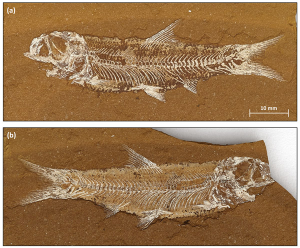 Cavenderichthys talbragarensis is the most abundant fossil fish in the Talbragar Fish Bed. The fish presents as a typical split fossil with part (a) and counterpart (b). During fossilisation, the original bone material was replaced by minerals (mostly quartz). Photos: Dr Michael Frese
"The methods I’ve been working on with my colleagues borrow techniques commonly used in other fields such as molecular biology or material science. Our work shows that fluorescence can be used to produce very detailed images of fossils from the Jurassic Talbragar Fish Bed [located 25km northeast of Gulgong, NSW] which is world-renowned for 150 million year old fossils.
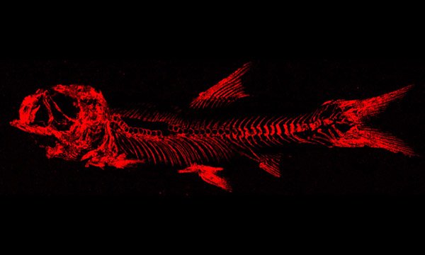
“We use ultra-violet light to image fossils," he said.
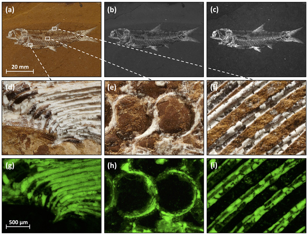
The equipment used by Dr Frese and his colleagues on a fossilised herring-like fish is commonly used to analyse DNA or fluorescent proteins in modern biology laboratories. The team also used x-ray fluorescence spectrometers to produce accurate elemental maps. If a fossil contains an element that is not present in the matrix (or vice versa), surprisingly accurate images can be produced – regardless of the colours present.
Minerals, such as quartz, often replace the original plant and animal remains during fossilisation. This phenomenon can be used for imaging if the minerals show fluorescence.
"The images we are producing allow us to see the tiniest details, things which until now would have remained a mystery lost to time," he said.
“For example, we can count the tiniest rays in the fin of a fish, which when looked at under a normal microscope just appear as a mass of white coloured material.
"Being able to see these details gives us far more information than we've ever had before. It gives us a better understanding of life millions of years ago and the processes which formed these fossils."
For this study, Dr Frese collaborated with Gerda Gloy from Bruker Nano Analytics, Dr Rolf Oberprieler from the CSIRO National Insect Collection, and Professor Damian Gore from Macquarie University.
The article Imaging of Jurassic fossils from the Talbragar Fish Bed using fluorescence, photoluminescence, and elemental and mineralogical mapping, detailing the techniques used in his study, has been published in the scientific journal PLOS ONE and is available online.